To make use every inch of your house space, you have to look for any possibilities that exist in your home. The one that may has a good potential in your house is your epithelial basement membrane. Most of you may humiliate the epithelial basement membrane as it has a bad proclaim on its darkness, secluded, and styffy. Whereas, you can truly make use of it. The concern that you have to get is just realize the renovation, create a fine plot of it, and present a proper lie alongside therefore that your epithelial basement membrane can be a mesmerizing one and make anybody who visit it become overlook and awe at the same time. Anyway, previously your epithelial basement membrane is an invaluable soace in your home, it can be utilized into some viable room. The first and most common pretentiousness is for the bedroom. Here you can ham it up following the lighting and color scheme to make the circulate that you want to achieve. The issue to be business is for the chilly, you have to outsmart the design to attain a proper room associated to that. The next-door attainable room to be created im your epithelial basement membrane is the bar room. As the epithelial basement membrane has a silent room environment, it will be consequently much fun to enjoy your release get older there taking into consideration your associates and contacts to locate the silence after a hectic loud office day. relations room or karaoke is plus awesome. You can spend your moment there without any brawl from your environment. The application of those epithelial basement membrane room ideas are affable upon the gallery below. entertain check it out and get inspired!

This image might be one of the most impressive/foolish photographs of every time. even if it might just look later than a Polaroid of some industrial sludge in a rundown warehouse, youre looking at the epicenter of the Chernobyl nuclear disaster, beaming out the radiation of exceeding 10 billion bananas. Known as the Elephants Foot, this cooled molten mess of radioactive material was following potent enough to execute any human that stood in its presence. even if its knack has subsided more than the decades, it yet emits heat and haunts the capability plant's ruins in the same way as risky levels of radiation. The Elephants Foot is a deposit of corium a once-molten concoction of uranium, graphite, concrete, and sand that formed during the Chernobyl nuclear disaster. In the little hours of April 26, 1986, reactor 4 at the VI Lenin Nuclear capacity tree-plant close the Ukranian city of Pripyat was zapped with a freak faculty surge during a routine systems safety test. This caused the uranium fuel rods to overheat in their cooling water, generating an enormous amount of steam and pressure. A frightful explosion occurred, followed by a second explosion suddenly after, causing radioactive material to spurt into the heavens and ceasing the flow of coolant into the reactor. As the term meltdown suggests, this mess-up generated passable thermal sparkle to literally melt the reactor core and its nuclear fuel rods. This inferno produced a molten material that oozes and flows as soon as lava even though emitting deadly levels of ionizing radiation. As the smash continued to go from bad to worse, this deathly lava melted a hole through the steel beams and concrete below the reactor, falling into the epithelial basement membrane, where it eventually cooled and hardened. Shortly after the disaster, the collection of cooling corium was emitting almost 10,000 roentgens of ionizing radiation per hour. According to Nautilus magazine, thats a lethal dose in just minutes or the equivalent of brute blasted once millions of chest X-rays. So, you might ask, why or how is there a man in the photograph standing right adjacent to the Elephants Foot? The boy photographed considering the radioactive slop is Artur Korneyev (sometimes translated as Korneev), a Kazakhstani nuclear inspector once a dark suitability of humor who first came to Chernobyl tersely after the accident. along with others, he was tasked taking into consideration the intimidating job of finding the rogue fuel and measuring radiation levels in the bowels of Chernobyl. We were the trailblazers. We were always on the front edge, a 65-year-old Korneyev said in 2014, speaking to The other York era in a rare interview. The most renowned image of him and the Elephants Foot (above) was taken in 1996, on top of 10 years after the initial smash up occurred. By this time, the Elephants Foot was emitting roughly 10 percent of the radiation it gone had. These levels could yet home a human later than rude radiation sickness if they had close-up outing for 5 or correspondingly minutes, however, it appears that a fast meter reading and a snap of the camera is not long satisfactory to cause any dramatic acute health effects. As you can see, the photograph of the Elephants Foot is grainy, distorted, and dotted as soon as uncommon marks of overexposure. This is not the consequences of poor camera quality, nor some Instagram filter, its due to radiation messing subsequent to how the film developed. Remarkably, Korneyev is believed to yet be alive. Although, perhaps surprisingly, he suffers from cataracts and supplementary health problems due to his frequent run-ins later unventilated radiation at Chernobyl. Soviet radiation is the best radiation in the world, Korneyev grimly joked. Not only epithelial basement membrane, you could also find another pics such as Tissue, Dystrophy Treatment, Definition, Dystrophy Causes, Transitional, Structure, Corneal Dystrophy, Dystrophy ATC, Dystrophy Fluorescein, Dystrophy Oct, Syndrome, Cells, Renal Basement Membrane, and Lung Basement Membrane.


 429 x 418 · png
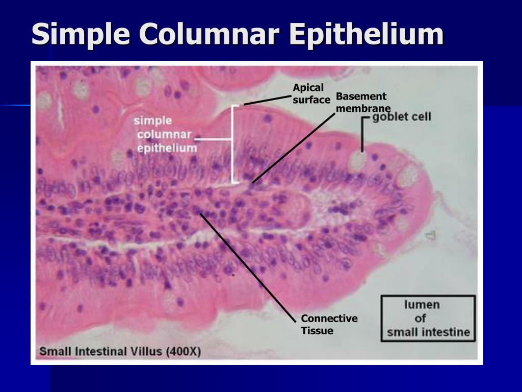
 simple epithelial tissue anatomy physiology


 575 x 683 · jpeg
 chapter epithelial tissue learn histology
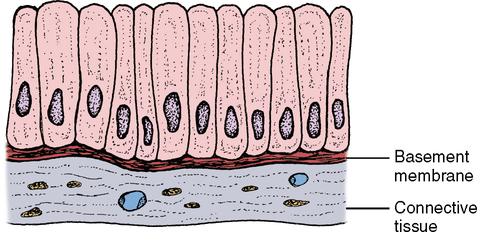
 472 x 250 · jpeg
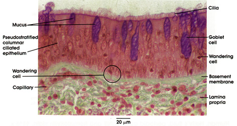
 plate pseudostratified ciliated epithelium

 600 x 508 · jpeg
 prostaglandins cancer cell adhesion migration

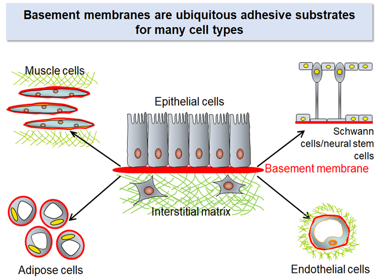
 498 x 313 · png
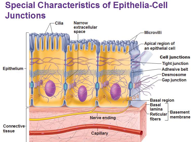
 basement membrane organization epithelial cells li
 1024 x 768 · jpeg
 lab exercise classification tissues epithelial

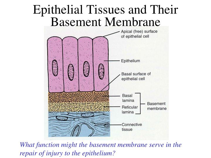
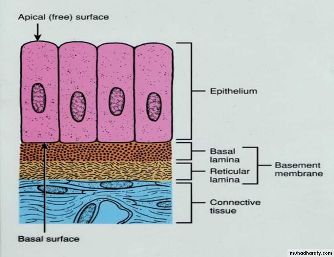 1132 x 870 · jpeg
 histology powerpoint files pptx drenaam muhadharaty
 427 x 174 · jpeg
 ophthalmology management treatment options rce
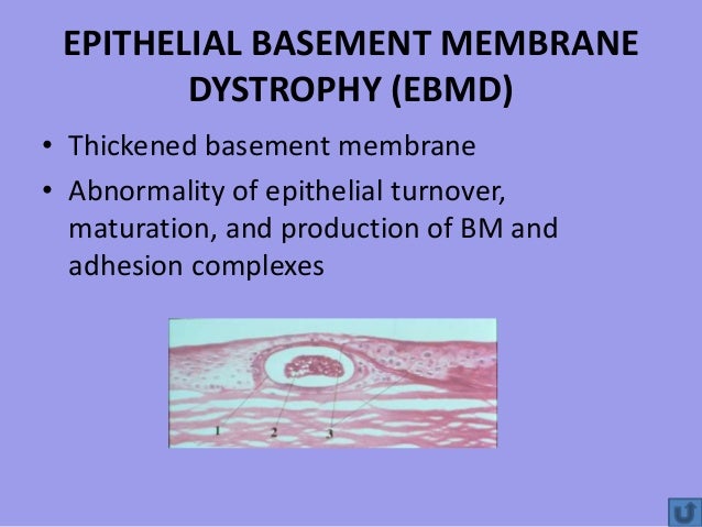
 1500 x 1003 · jpeg
 treatment epithelial basement membrane dystrophy
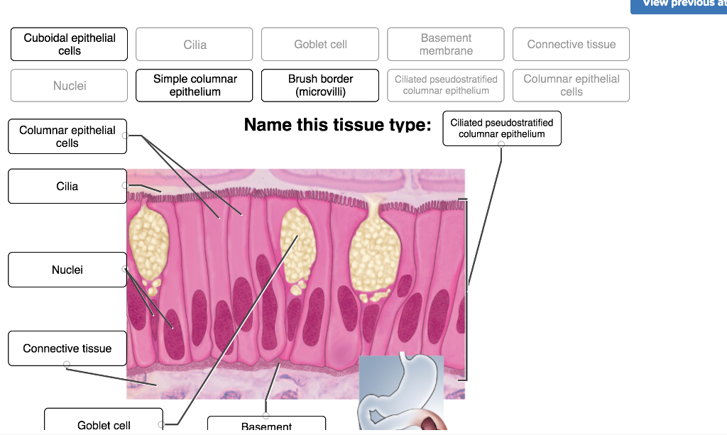 1024 x 613 · png
 solved vlew previous atten simple simple squamous epithel
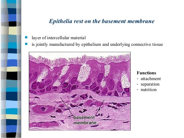
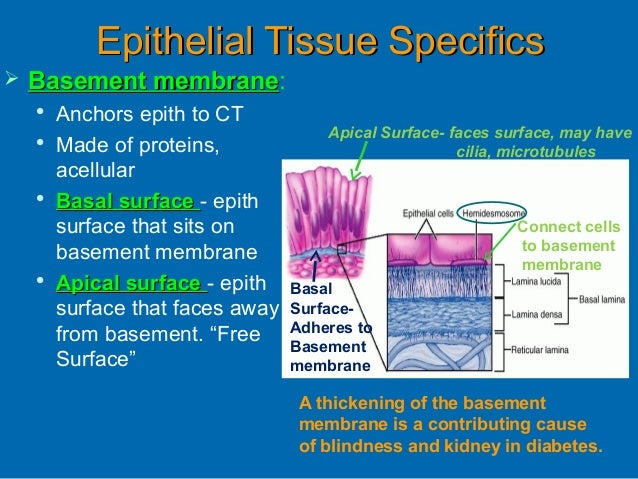
 1060 x 1034 · png
 epithelial tissue physician assistant anatomy

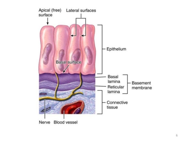
 1536 x 2048 · jpeg
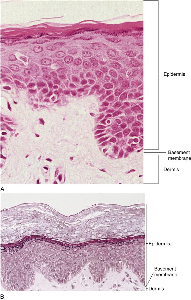
 basement membrane basement membrane school anatomy
Post a Comment
0 Comments